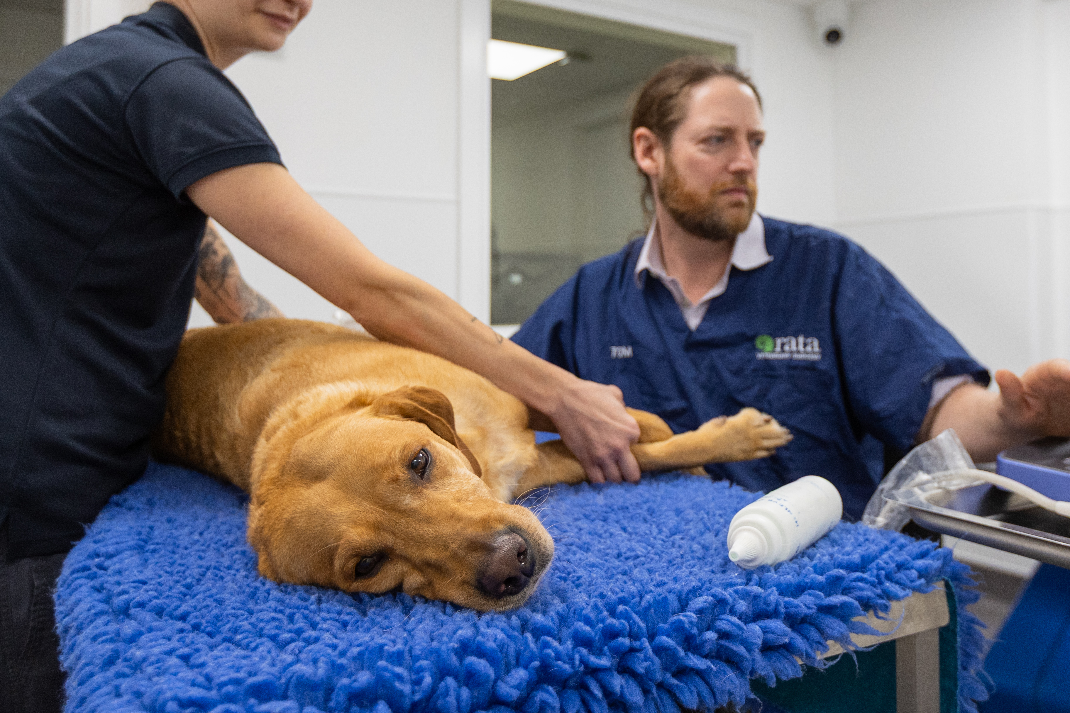
Thoracic ultrasound is a highly sensitive, non-invasive imaging tool that helps us assess your pet’s chest in more detail, particularly when X-rays alone are not conclusive.
It is especially useful for detecting fluid around the lungs, areas of lung consolidation (such as pneumonia), and abnormalities close to the chest wall. It also allows safe, real-time guidance for sampling fluid or masses when needed.
Common reasons for thoracic ultrasound include investigation of breathing difficulties, suspected pneumonia, fluid in the chest, chest masses or enlarged lymph nodes, and cases where further detail is needed after radiographs.
It is often used alongside X-rays to provide a more complete picture and help guide the most appropriate treatment for your pet.