

The Elbow joint
The elbow is a complex joint as it is composed of three bones – the Humerus (bone from shoulder to elbow), the Radius and Ulna (bones from elbow joint to wrist). In young dogs, the radius helps support the weight of the animal as it is transmitted through the humerus, whilst the ulna’s primary function is to allow smooth flexion and extension (bending and straightening) of the forelimb (front leg). In adult dogs, the load bearing is more equal between the radius and the ulna.
In a normal dog, these bones grow at exactly the same rate and are congruent (well aligned) with the radial head lining up with the Medial Coronoid Process (MCP) which is part of the ulna. This means they can articulate smoothly with each other to function as the elbow joint. Any articular (joint) surface is covered in cartilage which aids in this movement by protecting the bone surfaces.
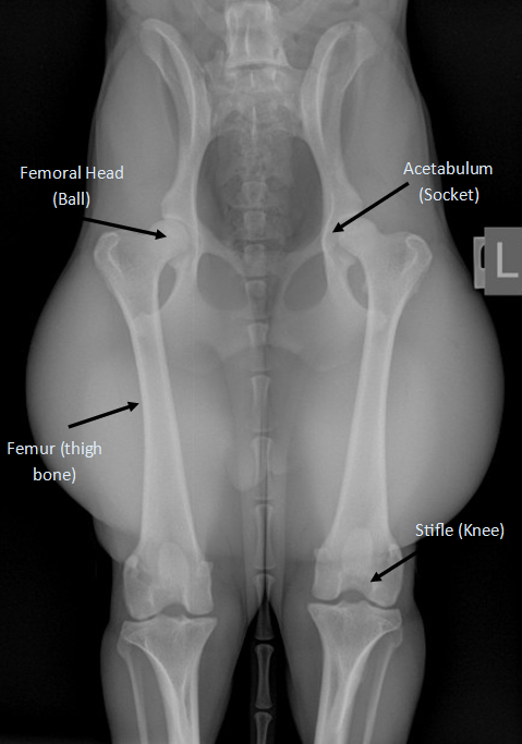
The Hip joint
The hip is composed of the femoral head (‘ball’ at the top of the thigh bone) and the acetabulum (‘socket’ in the pelvis). Muscles and ligaments help to keep this ball and socket joint in place. When the ball or socket are deformed the result is a partial dislocation of the hip joint when your pet is weight bearing. Over time this cause degeneration of the cartilage surfaces, remodelling of the bone, inflammation of the joint capsule and osteoarthritis.
Signs of joint dysplasia:
- Reluctance to exercise or lameness, usually onset is early in life (around 6-10 months). Pain and lameness may be seen later in life due to the development of osteoarthritis.
- Reduced range of motion in the joints
- Swelling or thickening of the elbow joint
- Muscle loss around the affected joint
Hip and Elbow Dysplasia

Bot of these conditions are complex in origin. Elbow dysplasia is a developmental problem relating to the incorrect development of the humerus, ulna and radius.The true cause of Hip dysplasia is unclear, however, it is accepted that HD is a result of a number of different genes as well as environmental influences
Both types of dysplasia lead to rubbing and extra pressure within the joint which cause cartilage erosion, fragmentation of bone and osteoarthritis.
Treatment of hip and elbow dysplasia:
This depends on the severity of the issue. Determining the best treatment can be challenging.
Some dogs will benefit from surgery whilst others require conservative management. All dogs with hip and elbow dysplasia (whether they have surgery or not) will develop arthritis as a result of the disease.
Diagnosis
An orthopaedic examination by your vet can help in the diagnosis of joint dysplasia. Our surgeons will check the joint for pain, range of motion and feel for any abnormal crepitus or popping in the joint.
Radiographs (x-rays) will be necessary to help in the diagnosis but frequently (in younger dogs), a CT scan will be required to provide further (more detailed information). Elbow arthroscopy is another useful diagnostic tool as it enables the surgeon to examine the joint directly by inserting a camera through a keyhole incision.
Outlook
This can be variable depending on the disease process and development of arthritis. Some dogs can be treated successfully with conservative treatment whilst others require surgery. A degree of stiffness and lameness (especially after exercise) is to be expected from dogs suffering with joint dysplasia.
Please view our information sheets on the different surgical techniques for more detailed information regarding expectations on recovery.
Useful Links:
How common is elbow dysplasia? HOW COMMON IS ELBOW DYSPLASIA? – YouTube
Canine elbow arthroscopy: a dog owner’s guide Canine elbow arthroscopy: A dog owner’s guide – YouTube
Subscribe to our newsletter
We won't share your details with any third parties